40 label the photomicrogram of the trachea
Labeled diagram of the lungs/respiratory system. - SERC View Original Image at Full Size. Labeled diagram of the lungs/respiratory system. Image 37789 is a 1125 by 1408 pixel PNG Uploaded: Jan10 14. Last Modified: 2014-01-10 12:15:34 A&P 2 Lab Unit 2 Flashcards | Quizlet Label the photomicrogram of the trachea. Identify these structures in the left-sided midsagittal view of the superior portion of the lower respiratory system. Identify the anatomical structures shown in the picture of the thorax. Identify the anatomical structures shown in a lateral view of the left lung.
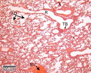
Anatomy A215 Virtual Microscopy - Indiana University Bloomington Anatomy A215 Virtual Microscopy Each alveolus is a small air space surrounded by an extensive capillary network. The epithelium which lines the alveoli is an extremely thin simple squamous in close proximity to the capillary walls. Alveoli make up the major part of the lung and give it its sponge-like appearance under the microscope.

Label the photomicrogram of the trachea
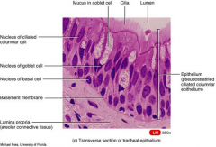
Histology, Alveolar Cells - StatPearls - NCBI Bookshelf Alveoli represent the most distal portion of the respiratory tract. There are approximately 500 million alveoli in the human body.[1] Each alveolus is separated from the other by an alveolar septum, which contains the pulmonary capillaries participating in gas exchange and connective tissue. BIO208 Lab Practical 2 - 10/6/2019 Lab Practical 2 Home - Course Hero 10/6/2019 Lab Practical 2 Question Label the structure with a "star" symbol beside it. 4 Incorrect Mark 0.00 out of 1.00 Answer: trachea The correct answer is: larynx (based on the document attached to the question as reference) Lab 2: Microscopy and the Study of Tissues - UW-La Crosse The lining of the trachea consists of a type of tissue called pseudostratified (ciliated) columnar epithelium. This single layer of ciliated cells appears stratified because the cells vary in their thickness and because their nuclei are located at different levels. 2 - Pseudostratified columnar epithelium (close-up view) Ciliated border
Label the photomicrogram of the trachea. Free Automated Malware Analysis Service - powered by Falcon Sandbox ... Submit malware for free analysis with Falcon Sandbox and Hybrid Analysis technology. Hybrid Analysis develops and licenses analysis tools to fight malware. Histology of trachea and lung - SlideShare 1. HISTOLOGY OF TRACHEA AND LUNG Dr.ushakannan,Asst.professor. 2. RESPIRATORY SYSTEM Conducting Part- responsible for passage of air and conditioning of the inspired air. Examples:nasal cavities,pharynx, trachea, bronchi and their intrapulmonary continuations. Respiratory Part-involved with the exchange of oxygen and carbondioxide between blood ... downloads.chartnettech.com o, o= +, o, o> (- o oI X 2Ð o? Ži 1 o‚ Þ , o@ Ü * ,Z† sS * sU * sM * sP * sc * se * s] * s_ * sg * s[ *.( € * {A * {B * {C * (' * (' *ž {D {E {D {F oG {H ... Label The Photomicrograph Of The Lung : 4 Chloro Dl Phenylalanine ... The lower respiratory system., put the following layers of the trachea in order from superficial to deep., label the structures of the upper respiratory . Label the photomicrogram of the lung segmental branch of pulmonary a. Relative amounts of glands, cartilage, smooth muscles and connective tissue fibers present in the wall of the tubes.
Trinidad State College Home Page Trinidad State is a Hispanic-Serving Institution (HSI) HSI is defined in federal law (the Higher Education Opportunity Act, Title V, 2008) as an accredited, degree-granting, public or private nonprofit institution of higher education with 25% or more total undergraduate Hispanic full-time equivalent (FTE) student enrollment. A&P 139 Chapter 19 Flashcards | Quizlet Label the photomicrogram of the lung. Label the photomicrogram of the trachea. Cricoid. Which of these laryngeal cartilages is single? Label these structures of the upper respiratory system. tidal volume. The volume of air that enters (or leaves) during a single respiratory cycle is the. (Get Answer) - Determine the angle of i, r and q this is reflection of ... Determine the angle of i, r and q this is reflection of light General Structure of Mucosa Label the structures that comprise the respiratory tract mucosa (mucous membrane). ... lung Segmental bronchus Trachea prey="" 11="" of="" 46="" next=""> Trachea histology of respiratory system low power Label the photomicrogram of the trachea. The Trachea (Human Anatomy): Picture, Function, Conditions, and More The trachea, commonly known as the windpipe, is a tube about 4 inches long and less than an inch in diameter in most people. The trachea begins just under the larynx (voice box) and runs down...
(Lee Ann C. Golper) Medical Speech-Language Pathol (BookFi) - Scribd [Lee_Ann_C._Golper]_Medical_Speech-Language_Pathol(BookFi).pdf - Free ebook download as PDF File (.pdf), Text File (.txt) or read book online for free. Medical Speech-Language Pathology: A Desk Reference, Third Edition ... write on label s.o.s. si opus si if necessary ss semis a half stat. statim immediately t.d.s. ter die sumendum to be taken three times daily t.i.d. ter in die three times a day t.i.n. ter in nocte three times at night ut dict ut dictum as directed v.i. vide infra see below via via by way of viz viz namely v.s. vide supra see above a Note: q.i.d ... The Bronchi: Anatomy, Function, and Treatment - Verywell Health At the bottom of the trachea is a ridge of cartilage called the carina. The carina essentially divides into the two primary bronchi; the right bronchi travels into the right lung and the left one to the left lung. Cartilage is what keeps the bronchi from collapsing during inhalation and exhalation. Solved Label the photomicrogram of the trachea. Cilia Lamina | Chegg.com Anatomy and Physiology questions and answers. Label the photomicrogram of the trachea. Cilia Lamina propria Submucosa Cilia Basement membrane Submucosa Epithelium Basement membrane Lamina propria Epithellum. Question: Label the photomicrogram of the trachea. Cilia Lamina propria Submucosa Cilia Basement membrane Submucosa Epithelium Basement ...
110 best Histology - Respiratory images on Pinterest | Lunges, Lungs ...
Can you label the lungs? Quiz - PurposeGames.com Printables and Stats. lungs. respiratory system. Labeling the Adult Skull Bones 16p Image Quiz. Body Planes 3p Image Quiz. 3p Image Quiz. Anterior Anatomical Body Regions 32p Image Quiz. Diagram of the Lower Limb 6p Image Quiz. Label the Small and Large Intestine. 8p Image Quiz.
The respiratory mucosa lines the conduction portion the air ...
Lab 2: Microscopy and the Study of Tissues - UW-La Crosse The lining of the trachea consists of a type of tissue called pseudostratified (ciliated) columnar epithelium. This single layer of ciliated cells appears stratified because the cells vary in their thickness and because their nuclei are located at different levels. 2 - Pseudostratified columnar epithelium (close-up view) Ciliated border

Photomicrograph Of Pseudostratified Ciliated Columnar Epithelium Of The ...
BIO208 Lab Practical 2 - 10/6/2019 Lab Practical 2 Home - Course Hero 10/6/2019 Lab Practical 2 Question Label the structure with a "star" symbol beside it. 4 Incorrect Mark 0.00 out of 1.00 Answer: trachea The correct answer is: larynx (based on the document attached to the question as reference)

Anatomy And Physiology Archive | October 25, 2017 | Chegg.com
Histology, Alveolar Cells - StatPearls - NCBI Bookshelf Alveoli represent the most distal portion of the respiratory tract. There are approximately 500 million alveoli in the human body.[1] Each alveolus is separated from the other by an alveolar septum, which contains the pulmonary capillaries participating in gas exchange and connective tissue.
Respiratory: The Histology Guide
Post a Comment for "40 label the photomicrogram of the trachea"