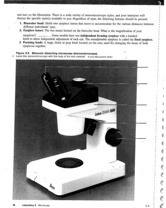
42 drag the labels onto the diagram to identify the parts of the microscope.
Solved cise 3 Review Sheet Art-labeling Activity 1 Γαιι ... - Chegg.com cise 3 Review Sheet Art-labeling Activity 1 Γαιι και Drag the labels onto the diagram to identify the parts of the microscope. Reset Hel rotating nose piece objective lenses mechanical stage coarso adjustment knob condenser | condensor knob DID NO ocul lenses substage light fine adjustment knob iris diaphragm lever onto on labels left the the the Drag diagram [X3ZR4V] Jun 21, 2019 · Parta drag the labels onto the diagram to identify structures and functions of the cardiovascular system. It is much better than the Drag and Drop onto Image. Drag the labels onto the diagram to identify the stem cells and stages of white blood cell and platelet production. Drag each label to the correct location on the chart.
the left onto the diagram labels on the Drag [VX2ZYQ] Follow these steps to set up a callout: Drag a custom callout shape from the Process Annotations stencil onto the drawing page. Draw a diagram and label all forces acting on the box. 3. With the drag-and-drop tool, just move the elements you like from the left side panel onto your page. Figure 5.

Drag the labels onto the diagram to identify the parts of the microscope.
a&p lab 3 hw Flashcards | Quizlet Drag the labels onto the diagram to identify the parts of the compound microscope (1 of 2). (figure 3.1) left column: arm mechanical stage right column: ocular lense ... Recall from the video the parts of a typical compound microscope. Drag the labels to identify the parts of the compound microscope. Not all labels will be used. 1. left column ... The Analysis of Biological Data, Second Edition - Academia.edu The Analysis of Biological Data by Michael C. Whitlock and Dolph Schluter Second Edition (z-lib.org) PDF Microscope quiz labeling - fusorebumobem.weebly.com Drag and drop the text labels onto the microscope diagram. All microscopes share features in common. In this interactive, you can label the different parts of a microscope. Use this with the Microscope parts activity to help students identify and label the main parts of a microscope and then describe their functions. Drag and drop the text ...
Drag the labels onto the diagram to identify the parts of the microscope.. Label the diagram to identify the organ systems Part A Drag the labels ... Part A Drag the labels onto the diagram to identify the anterior anatomical landmarks on the inferior half of the body. ANSWER: HelpReset Cephalic (head) Cervical (neck) Thoracic (chest) Brachial (arm) Abdominal (abdomen)Antecubital (front of elbow) Carpal (wrist) Manual (hand) 2/18/18, 10 (05 PMChapter 01 Homework Page 8 of 16Correct Art ... Ch 13 lab Map, Ch 12 lab map, CH 11 Lab MAP, Ch 10 lab map ... - Quizlet Identify the adult human skull structure labeled "C". [Be prepared to identify all labeled skull structures in this image on upcoming exams] lambdoid suture Identify the connective tissue proper cellular component labeled "F". fibroblast Drag the labels onto the diagram to identify the parts of the male reproductive system. look at pic LAB A&P Flashcards | Quizlet transverse Label the sectional planes of anatomical study. Label the diagram to identify the organ systems. Identify the quadrant that contains most of the stomach.. left upper quadrant When standing, moving toward the cranium is moving in __________ direction. a superior Drag the labels onto the diagram to identify the abdominopelvic regions. Solved Drag the labels onto the diagram to identify the - Chegg Expert Answer 100% (5 ratings) 1- ocular lens 2-rotating nose piece 3- condenser 4 … View the full answer Transcribed image text: Drag the labels onto the diagram to identify the parts of the microscope.
Bio2514 Week 3 The Microscope - Lab Topic.docx - Bio2514 Week 3 The ... Drag the labels onto the diagram to identify the parts of the compound microscope (1 of 2). Arm ocular lens Mechanical stage rotating nose piece Stage Objective lenses Condenser Iris diaphragm lever 3. The microscope slide rests on the __________ while being viewed. Stage 4. Your lab microscope is parfocal. Solved Drag the labels onto the diagram to identify the - Chegg Question: Drag the labels onto the diagram to identify the parts of the pelvis of the adult male. Reset Acetabulum Obturator foramen Sacroiliac joint llium Sacrum Pubis Ischium Pubic tubercle sma 100 Iliac crest Publc symphysis This problem has been solved! See the answer Show transcribed image text Expert Answer 100% (3 ratings) Compound Microscope Labeled Diagram - Quizlet QUESTION. The total magnification of a specimen being viewed with a 10X ocular lens and a 40X objective lens is. 15 answers. QUESTION. a mosquito beats its wings up and down 600 times per second, which you hear as a very annoying 600 Hz sound. if the air outside is 20 C, how far would a sound wave travel between wing beats. 2 answers. Drag the labels onto the diagram to identify the stages in which the ... Q: A microscope has a 12.0X eyepiece and a 59.0X objective lens 20.0 cm apart. Calculate (a) The total magnification, (b) The focal length of each lens, and (c) Where the object must be for a normal relaxed eye to see it in focus. Posted 2 days ago.
16 Parts of a Compound Microscope: Diagrams and Video Once you have an understanding of the parts of the microscope it will be much easier to navigate around and begin observing your specimen, which is the fun part! The 16 core parts of a compound microscope are: Head (Body) Arm Base Eyepiece Eyepiece tube Objective lenses Revolving Nosepiece (Turret) Rack stop Coarse adjustment knobs (Get Custom Answer) Identify The Nerves Of The Cervical Plexus Drag the labels onto the diagram to identify the nerves of the cervical plexus. Reset Help Hypoglossal nerve (XII) Transverse cervical nerve ADT WEEN Great auricular nerve III Phrenia nerve III Accessory nerve (X1) Supraclavicular nerves Using the figure above, match the number with the correct part of the microscope Cv1 A Fine and course adjustment knobs E2 B. Arm 13 c. Nosepiece FV4 D ... Chapter 8 Homework Test Questions - StudyHippo.com Looking through a light microscope at a dividing cell, you see two separate groups of chromosomes on opposite ends of the cell. ... Drag the labels onto the diagram to identify the stages of the life cycle. (Drag only blue labels onto blue targets and pink labels onto pink targets.) answer. question. The function(s) of meiosis is/are _____. answer. Parts of a microscope with functions and labeled diagram Q. List down the 18 parts of a Microscope. 1. Ocular Lens (Eye Piece) 2. Diopter Adjustment 3. Head 4. Nose Piece 5. Objective Lens 6. Arm (Carrying Handle) 7. Mechanical Stage 8. Stage Clip 9. Aperture 10. Diaphragm 11. Condenser 12. Coarse Adjustment 13. Fine Adjustment 14. Illuminator (Light Source) 15. Stage Controls 16. Base 17.

8 Best Images of Lens Diagram Worksheet - Microscope with Labeled Parts ...
Microscope Parts and Functions First, the purpose of a microscope is to magnify a small object or to magnify the fine details of a larger object in order to examine minute specimens that cannot be seen by the naked eye. Here are the important compound microscope parts... Eyepiece: The lens the viewer looks through to see the specimen.
31 Label The Indicated Parts Of The Microscope - Labels Database 2020
Simple Columnar Epithelium: A Labeled Diagram and Functions These form a brush border. They also increase the absorptive surface area of these cells. On a concluding note, simple columnar epithelium has two primary functions of absorption and secretion. In the small intestine, it facilitates the absorption of nutrients. It also secretes mucus, which helps to lubricate, moisten, and protect the surface.

Plant and Animal Cells - Labeled Graphics
Label the microscope - Science Learning Hub Use this with the Microscope parts activity to help students identify and label the main parts of a microscope and then describe their functions. Drag and drop the text labels onto the microscope diagram. If you want to redo an answer, click on the box and the answer will go back to the top so you can move it to another box.

35 Microscope Diagram To Label - Labels Information List
Solved Drag the labels onto the diagram to identify the - Chegg 93% (15 ratings) Transcribed image text: Part A Drag the labels onto the diagram to identify the parts of the corticospinal pathway. Reset Help rary Motor Corte Cerebral peduncle Medulla oblongata Anterior corticospinal tract To Lateral corticospinal tract skeletal muscles Midbrain Το Spinal cord skeletal muscles Motor nuclei of cranial ...

Questions And Answers On Labeled/Unlebled Diagrams Of A Human Cell : 16 ...
Drag Each Image To The Phase Of Meiosis I It Depicts Drag the labels onto the diagram to identify the stages of the cell cycle. Drag the blue labels to the blue targets to identify the stage of. A cell with a diploid number of 20 undergoes meiosis. Drag the labels from the left to their correct locations in the concept map on the right. Here in these phase the chromosome are.
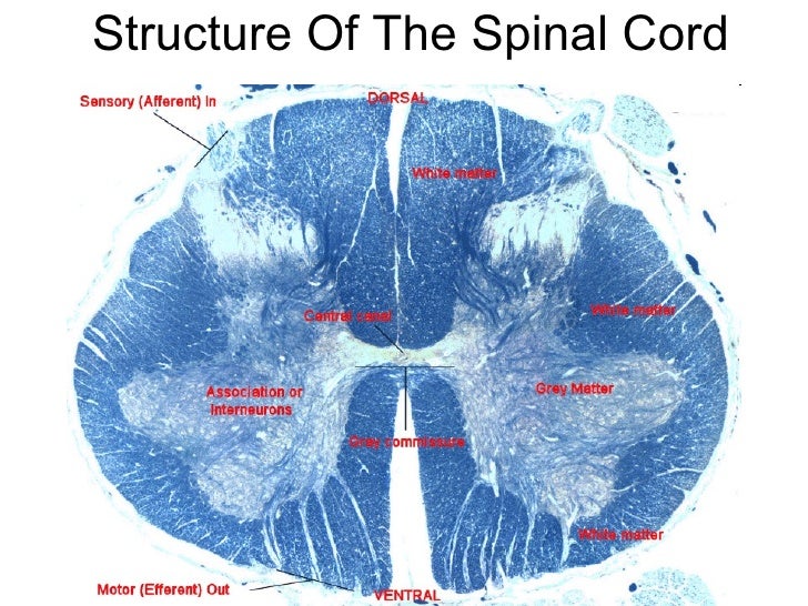
Neuroanatomy spinal cord
Label and identify the parts of a microscope? - Answers Best Answer. Copy. (Easier point view notes below) Eyepiece Lens: the lens at the top that you look through. They are usually 10X or 15X power. Tube: Connects the eyepiece to the objective lenses ...
Post a Comment for "42 drag the labels onto the diagram to identify the parts of the microscope."