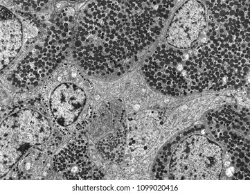
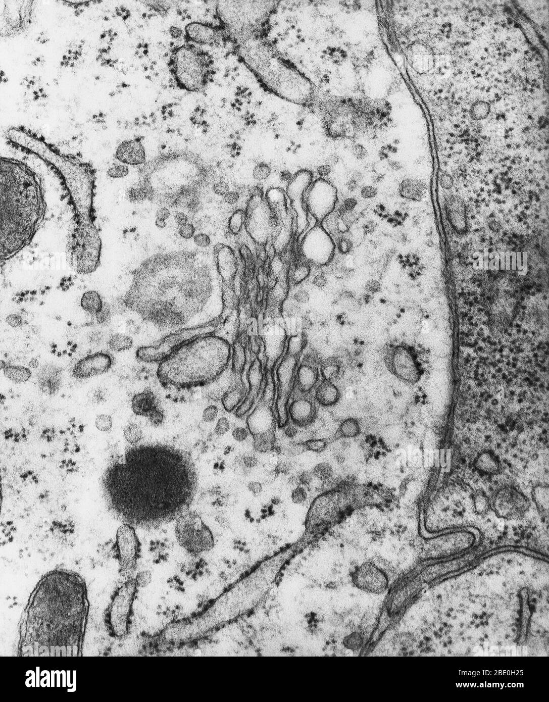
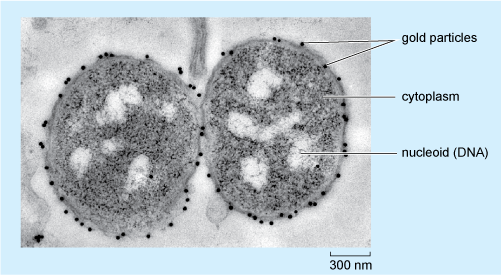
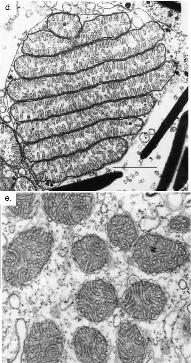
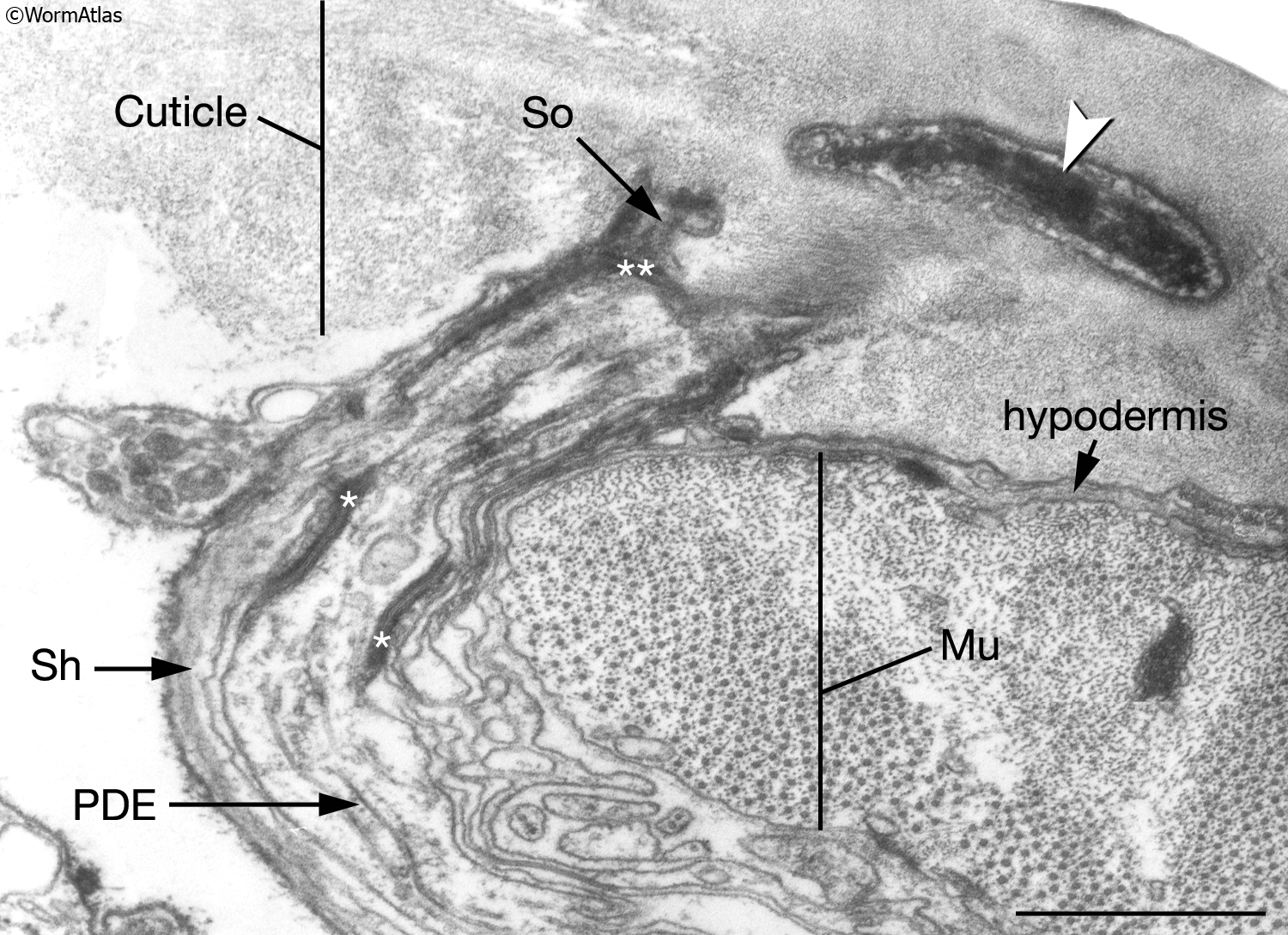
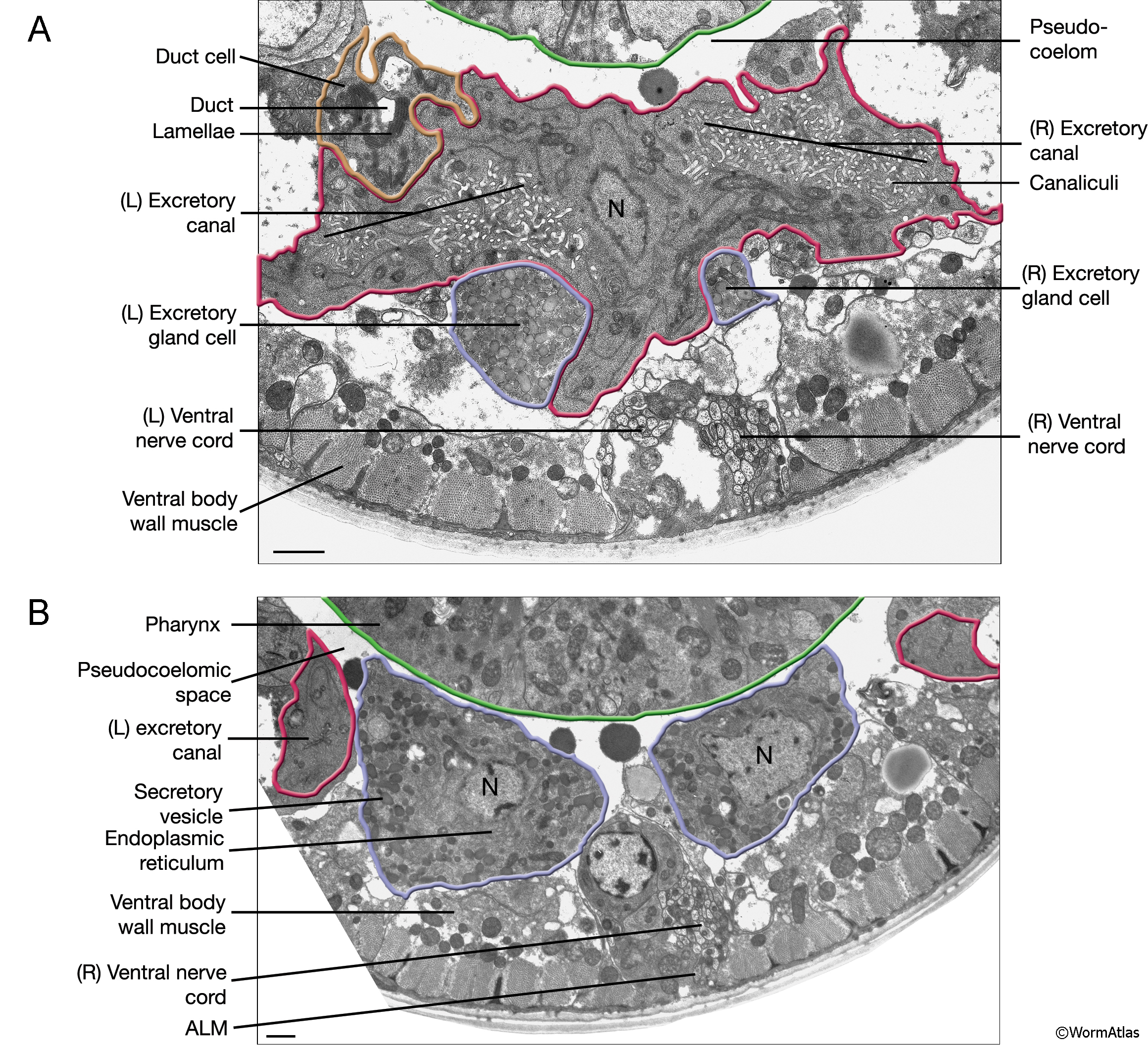
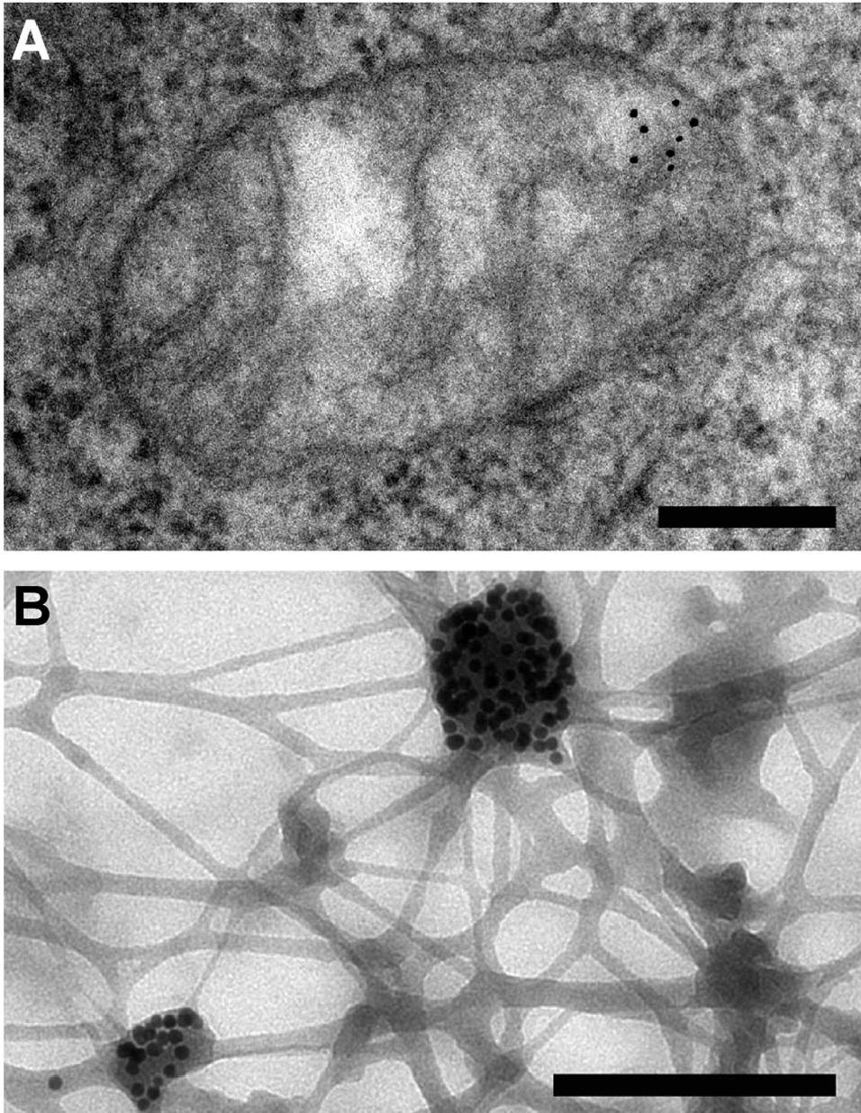
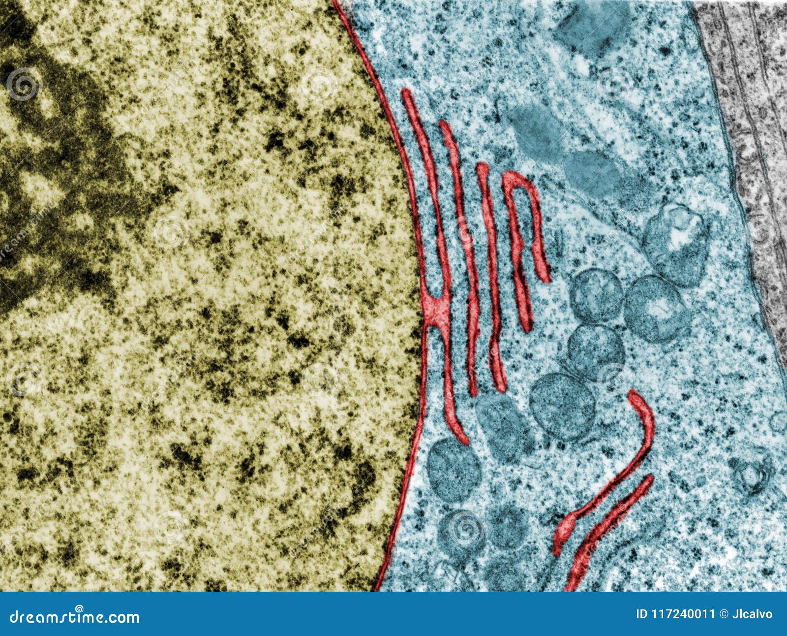
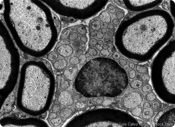
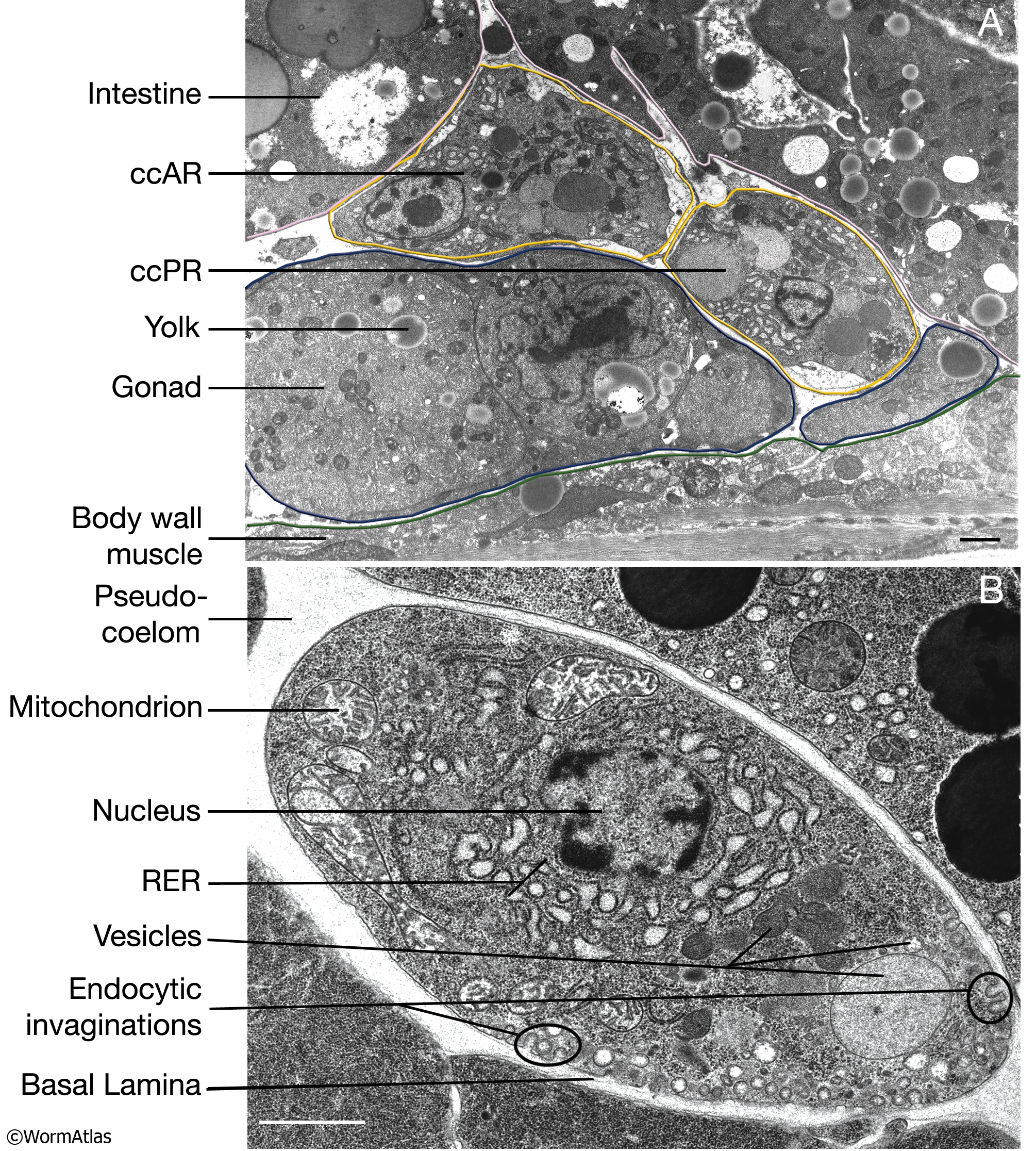
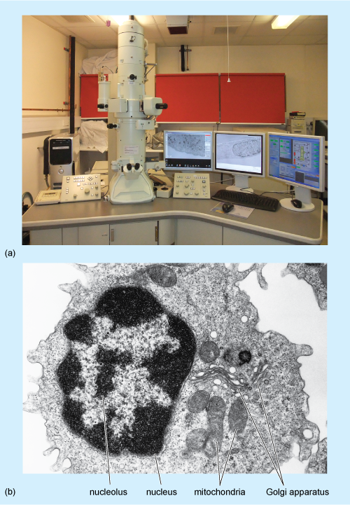
38 transmission electron micrograph labeled
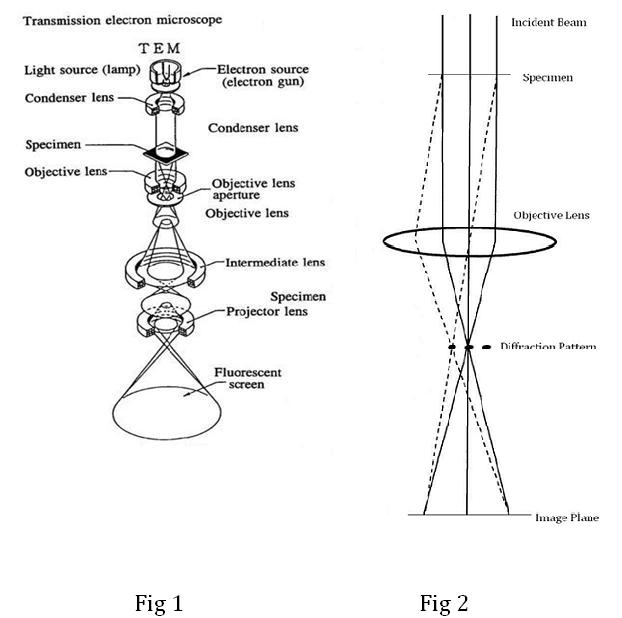
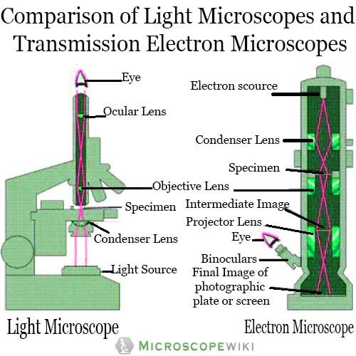
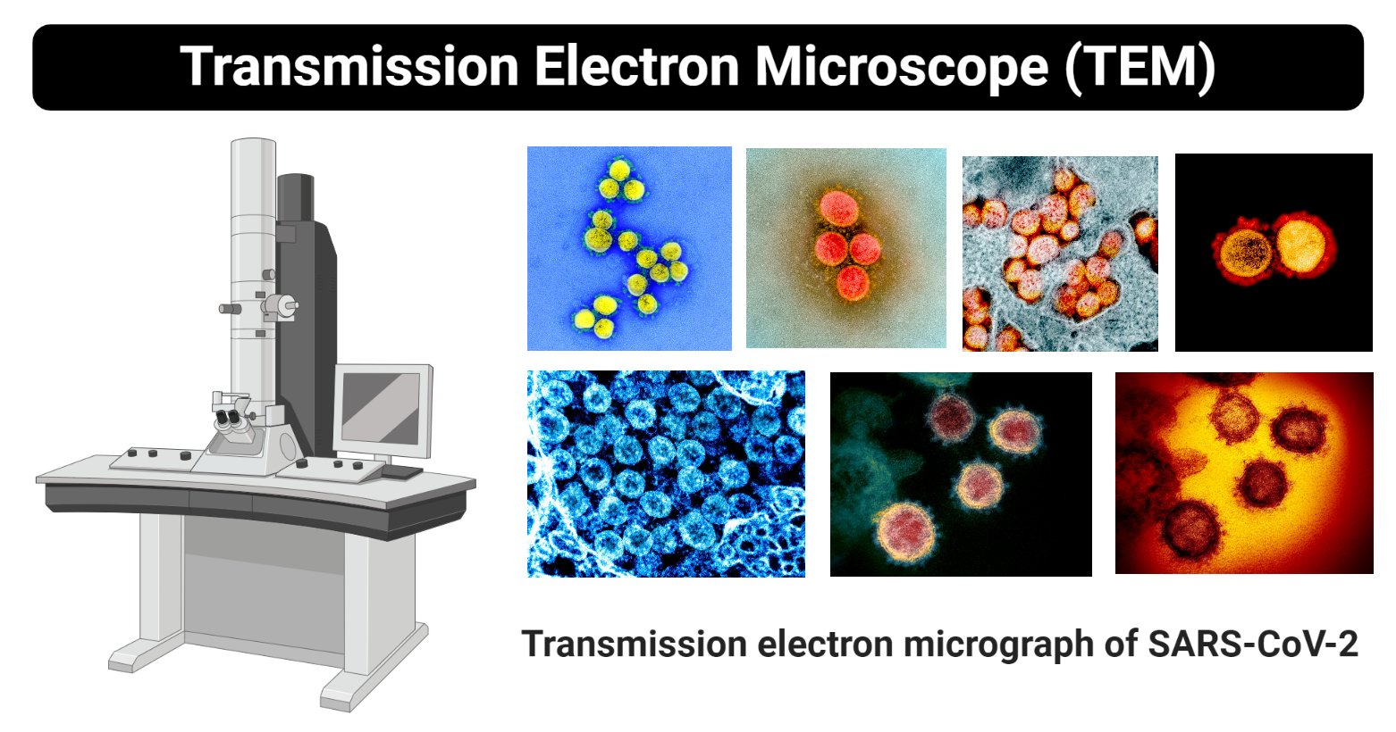
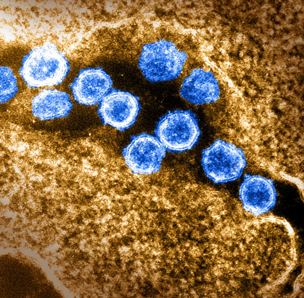
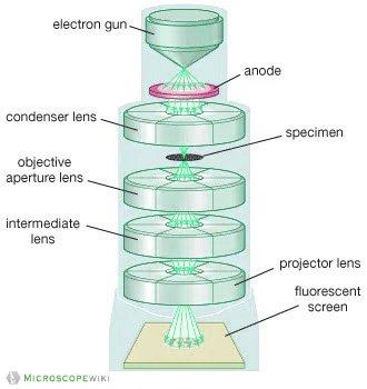
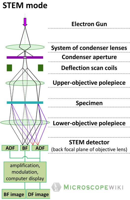
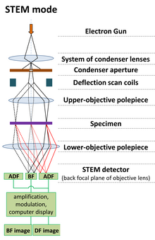
Transmission Electron Microscope (TEM)- Definition, Principle ... May 19, 2022 · Principle of Transmission Electron Microscope (TEM) The working principle of the Transmission Electron Microscope (TEM) is similar to the light microscope. The major difference is that light microscopes use light rays to focus and produce an image while the TEM uses a beam of electrons to focus on the specimen, to produce an image. Image Library | CDC Online Newsroom | CDC Note the spikes that adorn the outer surface of the virus, which impart the look of a corona surrounding the virion, when viewed electron microscopically. In this view, the protein particles E, S, and M, also located on the outer surface of the particle, have all been labeled as well.
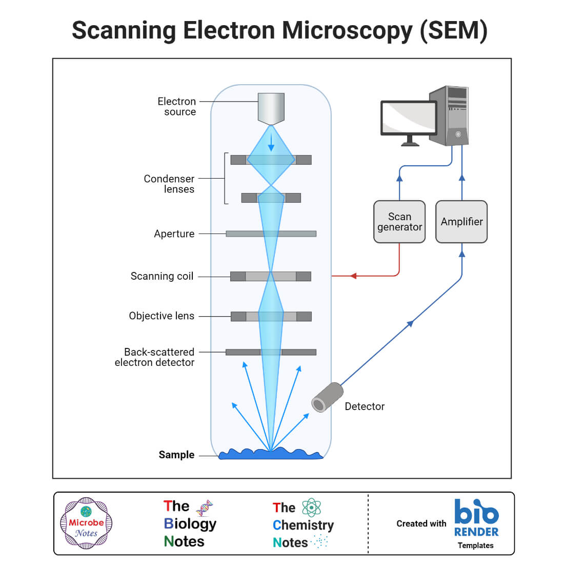
Scanning Electron Microscope (SEM)- Definition, Principle ... Mar 11, 2022 · The first Scanning Electron Microscope was initially made by Mafred von Ardenne in 1937 with an aim to surpass the transmission electron Microscope. He used high-resolution power to scan a small raster using a beam of electrons that were focused on the raster.
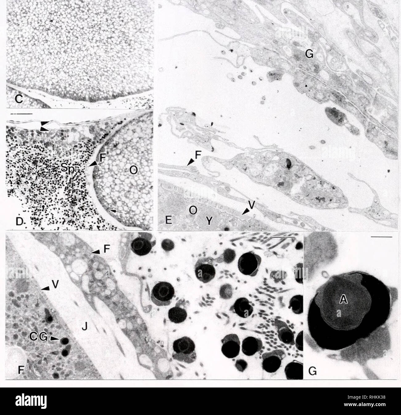
Transmission electron micrograph labeled
Electron microscope - Wikipedia An electron microscope is a microscope that uses a beam of accelerated electrons as a source of illumination. As the wavelength of an electron can be up to 100,000 times shorter than that of visible light photons, electron microscopes have a higher resolving power than light microscopes and can reveal the structure of smaller objects. Transmission Electron Microscopy - an overview ... 3.325.5.6 Transmission Electron Microscopy. TEM provides high-resolution imaging and is used for studying small areas or even single mineral platelets selectively. The TEM can be operated at different electron energies (often 100 keV for conventional TEM and 1 MeV for high-resolution imaging). The contrast of the TEM images is dependent on ... Fluorescence microscope - Wikipedia Fluorescence micrograph gallery A z-projection of an osteosarcoma cell, stained with phalloidin to visualise actin filaments. The image was taken on a confocal microscope, and the subsequent deconvolution was done using an experimentally derived point spread function.
Transmission electron micrograph labeled. Introduction to Pathogens - Molecular Biology of the Cell ... We then explore the wide variety of organisms that are known to cause disease in humans.Figure 25-1Parasitism at many levels(A) Scanning electron micrograph of a flea. The flea is a common parasite of mammals—including dogs, cats, rats, and humans. Fluorescence microscope - Wikipedia Fluorescence micrograph gallery A z-projection of an osteosarcoma cell, stained with phalloidin to visualise actin filaments. The image was taken on a confocal microscope, and the subsequent deconvolution was done using an experimentally derived point spread function. Transmission Electron Microscopy - an overview ... 3.325.5.6 Transmission Electron Microscopy. TEM provides high-resolution imaging and is used for studying small areas or even single mineral platelets selectively. The TEM can be operated at different electron energies (often 100 keV for conventional TEM and 1 MeV for high-resolution imaging). The contrast of the TEM images is dependent on ... Electron microscope - Wikipedia An electron microscope is a microscope that uses a beam of accelerated electrons as a source of illumination. As the wavelength of an electron can be up to 100,000 times shorter than that of visible light photons, electron microscopes have a higher resolving power than light microscopes and can reveal the structure of smaller objects.













Post a Comment for "38 transmission electron micrograph labeled"