41 how to label nmr spectra
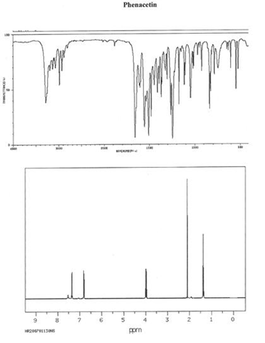
PDF MestreNova Quick Guide - Department of Chemistry The Full Spectrum icon will get the entire spectrum back in horizontal (ppm) and vertical (intensity) dimensions. The shortcut'F' will get the entire spectrum back in the horizontal (ppm) dimension.While the shortcut 'H' or icon will fit the tallest peak in the visible region to the top of the page. NMR Spectrum of Phenacetin | Thermo Fisher Scientific - US Compact, affordable bench-top NMR spectroscopy has never been easier. Chemical name: Phenacetin (N- (4-Ethoxyphenyl) acetamide) Concentration: 6% (w / w; 0.5 M) in CDCl3 CAS: 62-44-2 Field: 82 MHz Nuclear testing: 1 H Applications: Pharmaceuticals, forensics, bench analysis, productivity Back to February 2015 Tech Talk About NMR Tech Talk
Citrate | C6H5O7-3 - PubChem Lithium Citrate is the citrate salt of lithium, a monovalent cation with antimanic activity.Although the exact mechanism is unclear, lithium might exert its mood-stabilizing effect via reduction of catecholamine concentration mediated through transneuronal membrane transport of sodium ion by sodium-potassium-stimulated adenosine triphosphatase (Na-K-ATPase).

How to label nmr spectra
Isotopic Labeling for NMR Spectroscopy of Biological Solids The simplest and most cost-effective biosynthetic labeling method for protein solid-state NMR is to uniformly label all carbon and nitrogen atoms with 13 C and 15 N. In this way, a single protein sample can in principle provide all the structural constraints - dihedral angles and distances - about the protein. NMR Spectroscopy - Michigan State University To begin with, the nmr spectrometer must be tuned to a specific nucleus, in this case the proton. The actual procedure for obtaining the spectrum varies, but the simplest is referred to as the continuous wave (CW) method. A typical CW-spectrometer is shown in the following diagram. Sotalol: Uses, Interactions, Mechanism of Action - DrugBank Generic Name Sotalol DrugBank Accession Number DB00489 Background. Sotalol is a methanesulfonanilide developed in 1960. 6 It was the first of the class III anti arrhythmic drugs. 6 Sotalol was first approved as an oral tablet on 30 October 1992. 8 A racemic mixture of sotalol is currently formulated as a tablet, oral solution, and intravenous injection indicated for life threatening ...
How to label nmr spectra. PDF NMR Guidelines for ACS Journals - American Chemical Society H NMR spectrum should arise from the normally compound, not the solvent. 2.4 All peaks in the 1 H NMR spectrum should be integrated. Chemical shift values should be included. 2.5 The solvent peak should be clearly labeled on the spectrum. 2.6 All peaks should be visible on the spectrum. Insets are encouraged to show expanded regions. 6.6 ¹H NMR Spectra and Interpretation (Part I) – Organic … Figure 6.6b The chemical shift scale in H NMR spectra For protons that are shielded , because of the B local caused by circulating electrons, the magnetic field experienced by the proton, B eff , is smaller than the applied external field, B o , so the protons have a lower resonance frequency and have smaller chemical shift values. How to Interpret Proton NMR Spectra - theSpectroscopy let's interpret the 1H NMR spectrum for a compound with the molecular formula C3H7Br. First, we observe that there are three distinct signals, with chemical shifts of approximately δ 3.4, 1.8, and 1.1. One of these signals (δ 3.4) is noticeably downfield of the others, indicating hydrogen atoms that are likely to be near an electronegative group. PDF ANALYSIS OF H NMR SPECTRA - University of Texas at Dallas spectrum to five signals, corresponding to 5 different sets of protons. 2. RELATIVE INTENSITIES OF THE PEAKS, OR SIGNALS. The term intensity, when used in reference to NMR signals, indicates the area under the peak. The areas under the peaks are given by an integrator when recording a spectrum.
NMR- drawing and labelling spectra. - The Student Room NMR- drawing and labelling spectra. I've been given the molecule PF2H and I have to sketch the phosporous, flourine and hydrogen NMR spectra. Then label the peaks and coupling constants. I think I've worked out the shape to be trigonal bipyramidal but I'm a bit confused about drawing the spectra. I think the way that I've drawn the shape shows ... Stacked Spectra - Mestrelab Resources Graphical selection of spectra. In order to be able to select a bunch of spectra graphically with the mouse, it is necessary to display the spectra in the stacked plot mode. To select a single spectrum, left click on the spectrum trace while keeping pressed the Alt key To deselect a spectrum: left click + Ctrl + Alt In order to select multiple ... Tauroursodeoxycholic acid | C26H45NO6S - PubChem Tauroursodeoxycholic acid is the more hydrophilic form of ursodeoxycholic acid, which is the more abundant naturally produced bile acid in humans.Tauroursodeoxycholic acid, on the other hand, is produced abundantly in bears and has been used for centuries as a natural remedy in some Asian countries. How to reference 1D and 2D NMR spectra? … Part 2 - ACD/Labs In the case where there are multiple choices for a reference point, I typically lean towards an intense singlet as the 2D counterpart tend to be easy to locate on a 2D NMR spectrum. Checking the COSY data, the diagonal correlation at 5.9, 5.9 ppm is properly aligned to the 1D resonance at 5.9 ppm. Therefore, no further data manipulation is needed.
Interpreting C-13 NMR Spectra - Chemistry LibreTexts The 13 C NMR spectrum for ethanol The NMR spectra on this page have been produced from graphs taken from the Spectral Data Base System for Organic Compounds ( SDBS) at the National Institute of Materials and Chemical Research in Japan. Remember that each peak identifies a carbon atom in a different environment within the molecule. 13C Carbon NMR Spectroscopy - Chemistry Steps In general, when you start analyzing a 13 C NMR, split the spectrum in two parts by drawing a line at 100 ppm; below this value you have the saturated functional groups, and beyond that is the unstructured region. Saturated carbon atoms connected to electronegative heteroatoms give signal from 30-90 ppm. NMR and IR spectra predictions in ChemDraw and Chem3D ChemDraw and Chem3D have several tools for predicting NMR (Nuclear Magnetic Resonance) spectra and IR (Infrared) spectra . This article will discuss the key features and methods for NMR and IR prediction. This article has an associated webinar that provides examples and has downloads of the molecules used, allowing a user to personally try out the NMR and IR predictions. Nuclear magnetic resonance spectroscopy - Wikipedia Nuclear magnetic resonance spectroscopy, most commonly known as NMR spectroscopy or magnetic resonance spectroscopy (MRS), is a spectroscopic technique to observe local magnetic fields around atomic nuclei. The sample is placed in a magnetic field and the NMR signal is produced by excitation of the nuclei sample with radio waves into nuclear magnetic resonance, …
Please sketch out the theoretical 1H-NMR spectrum for your ...
Lifestyle | Daily Life | News | The Sydney Morning Herald The latest Lifestyle | Daily Life news, tips, opinion and advice from The Sydney Morning Herald covering life and relationships, beauty, fashion, health & wellbeing

How2: Interpret a proton NMR spectrum
Isotopic Labelling | Protein NMR This page describes some of the different isotopic labelling strategies that are commonly used in Protein NMR. In most cases (except in cell-free labelling), the protein is expressed by bacteria. The isotopic labels are introduced by feeding the bacteria specific nutrients.
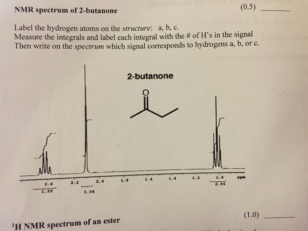
Solved NMR spectrum of 2-butanone (0.5) Label the hydrogen ...
Deciphering 1H NMR Spectra - StudyOrgo.com Once a NMR spectrograph is recorded, 4 pieces of information can be determined from the data as long as the chemical formula of the compound is known. To illustrate the points, we will consider the following 1H-NMR spectrum of the C 5 H 10 O. Signal Count - Number of unique hydrogens This is the easiest to interpret.
Label-free quantitative 1H NMR spectroscopy to study low ...
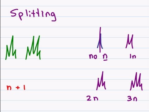
How to Read NMR Spectra of Organic Compounds | Study.com If a chemically distinct H atom sees N identical H atoms bonded to a neighboring C atom (s), the original peak will split into a set of 'N + 1' peaks in the spectrum. We call this the N + 1 rule,...
5.7: 13C-NMR Spectroscopy - Chemistry LibreTexts
Principles of NMR: How to Read Spectra and Couplings | Hatsudy Simply put, you can think of NMR as a method to investigate the electron density around a hydrogen atom (proton). By checking the spectra, you can infer the structural formula of the compound. For low and high magnetic fields, for example, the following NMR spectra are displayed.
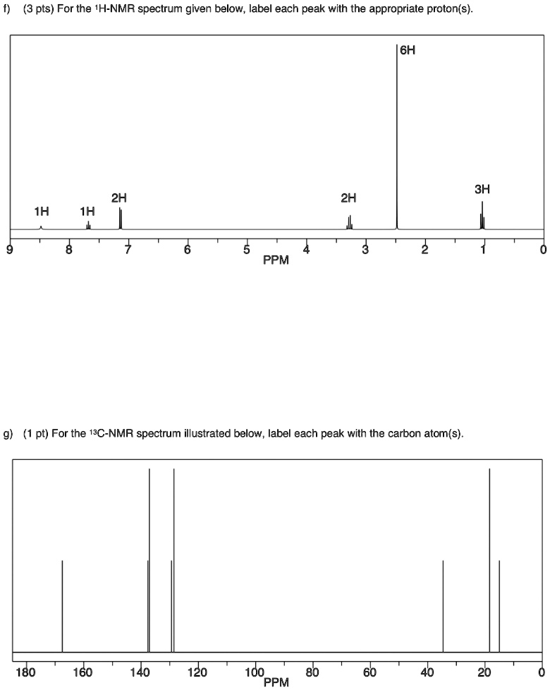
SOLVED: pts) For the 'H-NMR spectrum given below; label each ...
Prednisolone acetate | C23H30O6 - PubChem Prednisolone Acetate is the acetate salt form of prednisolone, a synthetic glucocorticoid with anti-inflammatory and immunomodulating properties.As a glucocorticoid receptor agonist, prednisolone acetate binds to specific intracellular glucocorticoid receptors, and causes the ligand-receptor complex to be translocated to the nucleus where it initiates the transcription of glucocorticoid ...
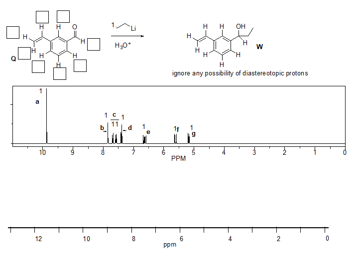
OneClass: 24. Match the label the protons on the compound Q ...
2-(2-Aminoethoxy)ethanol | C4H11NO2 - PubChem 2-(2-aminoethoxy)ethanol appears as a colorless liquid with a faint fishlike odor. Combustible but difficult to ignite. Corrosive to tissue. Combustion produces toxic oxides of nitrogen.
Solved] Identify the structure from the NMR spectrum and ...
Digital micelles of encoded polymeric amphiphiles for direct … 03/11/2022 · The 1 H NMR spectra recorded for polyurethanes exhibited characteristic resonance signals ascribed to carbamate protons (∼ 9.6 ppm) and methylene protons of benzyl moieties (4.8–5.6 ppm; Fig ...

Proton nuclear magnetic resonance - Wikipedia
NMR - Interpretation - Chemistry LibreTexts Here is the general strategy for solving structure with NMR: Molecular formula is determined by chemical analysis such as elementary analysis Double-bond equivalent (also known as Degree of Unsaturation) is calculated by a simple equation to estimate the number of the multiple bonds and rings.
Solved Label all the peaks for the H NMR spectrum for | Chegg.com
PDF NMR of Proteins - University at Buffalo • simple 152D 1H, N correlation NMR spectra of proteins • reduce complex spectra to simple ones based on isotope editing • reduce/eliminate spectral overlap/spectral degeneracy • correlate amide 1H-15N pairs • this spectrum demonstrates 1). that you can express your protein, 2). That you can isotopically label your protein, 3). That your
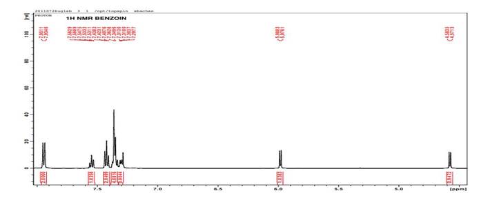
Solved) - Interpret the 1H and 13C NMR spectras of Benzoin ...
Nuclear magnetic resonance (NMR) spectroscopy: Hydrogen The NMR spectrum of ethyl benzene, C 6 H 5 CH 2 CH 3, is shown below.The frequencies correspond to the absorption of energy by 1 H nuclei, which are protons. Notice that there are three major peaks of differing heights. Ethyl benzene 1 H NMR spectrum Each peak corresponds to a hydrogen atom in a different molecular environment.
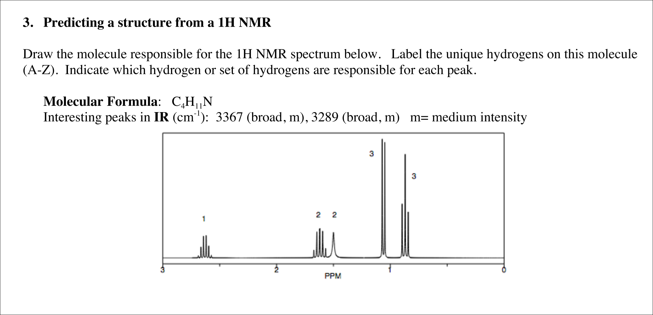
Solved Draw the molecule responsible for the 1H NMR spectrum ...
How To Label NMR Spectra - YouTube How To Label NMR Spectra 2,688 views Aug 31, 2020 9 Dislike Share Save Tyler Parra 309 subscribers Show more General Chemistry 1 Review Study Guide - IB, AP, & College Chem Final Exam The Organic...

1 H NMR spectrum of 10 in the downfield region in CDCl 3 (for ...
NMR spectroscopy - An Easy Introduction - Chemistry Steps NMR spectroscopy is the most common and comprehensive technique for studying the structure of organic molecules. In a broad sense, it still works by the same principle as other spectroscopies, and that is the interaction of the molecule with certain type of energy to produce different energy states and deduce information based on these differences.

Label the signals due to ha, hb, and hc in the 1h nmr spectra ...
How to interpret NMR spectra - University of Texas at Austin 1.2 An NMR spectrum is a plot of absorbance versus frequency. 1.2A To make different spectra directly comparable, a standard is used for all NMR spectra. For 1H NMR spectra, the standard is called tetramethylsilane (TMS) and a small amount of TMS is usually added to any 1H NMR sample.
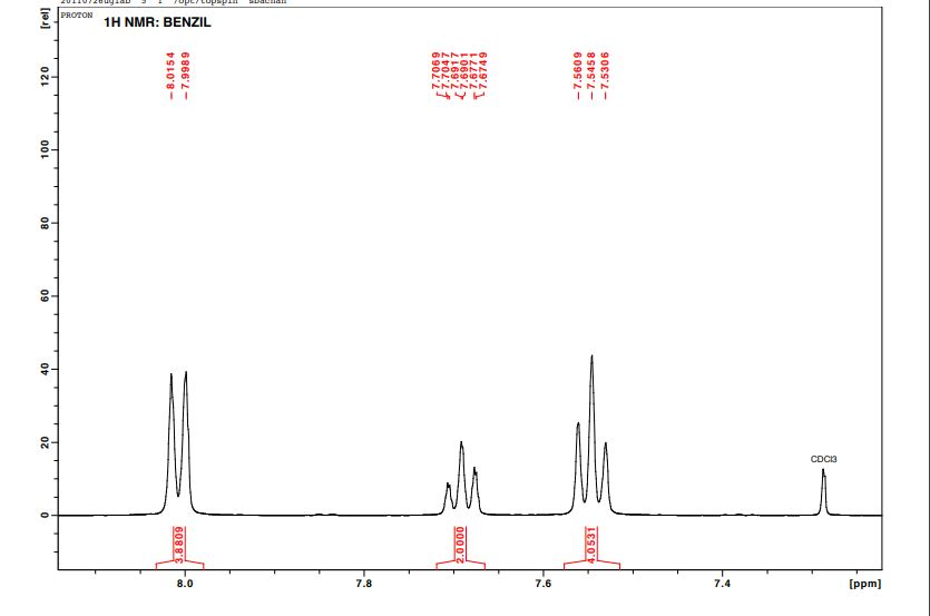
Solved Interpret both the 1 H and 13C NMR spectra of Benzil ...
How To Analyze The Peaks Of H-NMR Spectroscopy - YouTube Proton NMR - How To Analyze The Peaks Of H-NMR Spectroscopy - YouTube 0:00 / 11:31 Proton NMR - How To Analyze The Peaks Of H-NMR Spectroscopy 1,061,820 views Jan 23, 2013 9K Dislike...
NMR Spectroscopy
4.7: NMR Spectroscopy - Chemistry LibreTexts Aug 28, 2022 · Theory. The chemical theory that underlies NMR spectroscopy depends on the intrinsic spin of the nucleus involved, described by the quantum number S. Nuclei with a non-zero spin are always associated with a non-zero magnetic moment, as described by Equation \ref{1}, where μ is the magnetic moment, \(S\) is the spin, and γ is always non-zero.
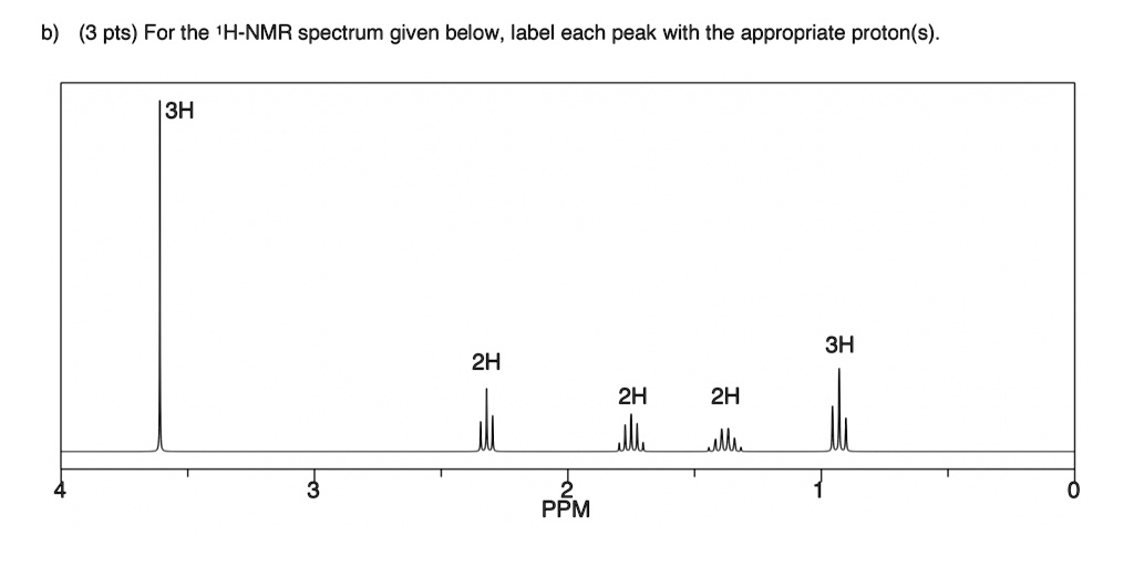
SOLVED: b) (3 pts) For the 'H-NMR spectrum given below; label ...
A Step-By-Step Guide to 1D and 2D NMR Interpretation - Emery Pharma Step 1: ¹H-NMR The first step in structural characterization is 1-dimensional proton ¹H-NMR. The chemical shift, multiplicity, coupling constants, and integration are all factors to consider when assigning protons. In this example, only three protons can be assigned by the proton spectrum alone: protons 3, 4, and 6.
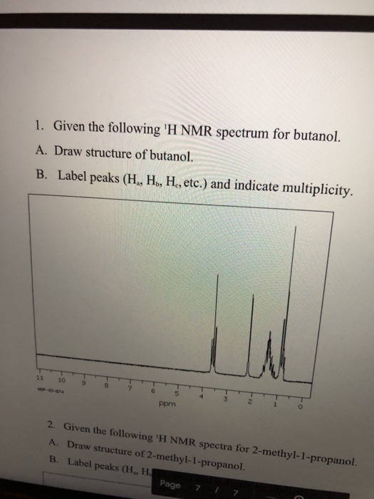
Solved 1. Given the following 'H NMR spectrum for butanol ...
The Basics of Interpreting a Proton (1H) NMR Spectrum - ACD/Labs 1 H NMR is the go-to technique to help identify or confirm the structure of organic compounds or those that contain protons. A solution-state proton spectrum is relatively fast to acquire, compared with other nuclei, and a lot of information about the structure of a compound can be deduced from it.
Solved) - Identify and analyze the spectral data for ...
NMR Spectrum of Aspirin | Thermo Fisher Scientific - US The 1 H NMR spectrum of a 4% (w/w; 330 mM) solution of aspirin in chloroform-D was measured at 82 MHz using the Thermo Scientific picoSpin 80 NMR spectrometer. Aspirin contains aliphatic, aromatic and carboxylic acid protons that span a wide range of the 1H spectrum, and signal integration reveals a 3:4:1 intensity distribution, respectively.
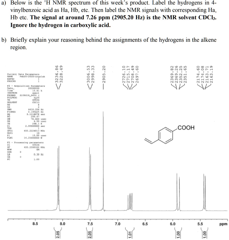
SOLVED: Below is the 'H NMR spectrum of this week's product ...
How to Identify Molecular Fragments from NMR and IR Spectra When trying to determine the structure of a compound based on its molecular formula, you can use nuclear magnetic resonance (NMR) and infrared (IR) spectroscopy to help you identify the fragments of the molecule. (Once you identify these fragments, you can identify the molecule's structure.)
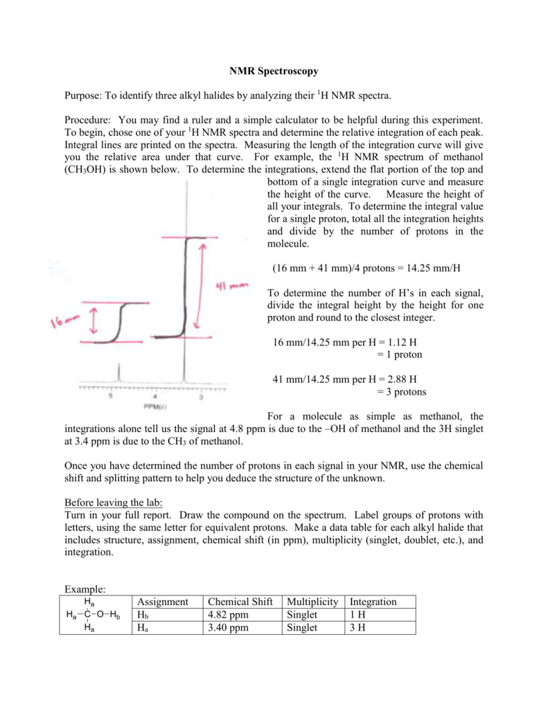
NMR Spectroscopy
Emergent charge order in pressurized kagome superconductor … 23/11/2022 · The NMR spectra around P c1 and P c2 show the characteristic structure for the coexistence of two distinct phases, suggesting a first-order-like quantum phase transition 47.
.jpg)
Using Benchtop Proton NMR Spectroscopy to Determine the Main ...
Sotalol: Uses, Interactions, Mechanism of Action - DrugBank Generic Name Sotalol DrugBank Accession Number DB00489 Background. Sotalol is a methanesulfonanilide developed in 1960. 6 It was the first of the class III anti arrhythmic drugs. 6 Sotalol was first approved as an oral tablet on 30 October 1992. 8 A racemic mixture of sotalol is currently formulated as a tablet, oral solution, and intravenous injection indicated for life threatening ...

13C Carbon NMR Spectroscopy - Chemistry Steps
NMR Spectroscopy - Michigan State University To begin with, the nmr spectrometer must be tuned to a specific nucleus, in this case the proton. The actual procedure for obtaining the spectrum varies, but the simplest is referred to as the continuous wave (CW) method. A typical CW-spectrometer is shown in the following diagram.
NMR - Interpretation - Chemistry LibreTexts
Isotopic Labeling for NMR Spectroscopy of Biological Solids The simplest and most cost-effective biosynthetic labeling method for protein solid-state NMR is to uniformly label all carbon and nitrogen atoms with 13 C and 15 N. In this way, a single protein sample can in principle provide all the structural constraints - dihedral angles and distances - about the protein.
NMR Guidelines for ACS Journals
1 H NMR spectra of NA4, Peak labels: A, 3,6 anhydrogalactose ...
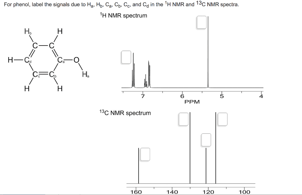
OneClass: For phenol, label the signals due to Ha, Hb, ca, cb ...
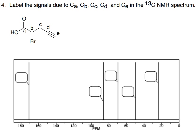
SOLVED: Label the signals due to Ca, Cb, Cc, Cd, and Ce in ...
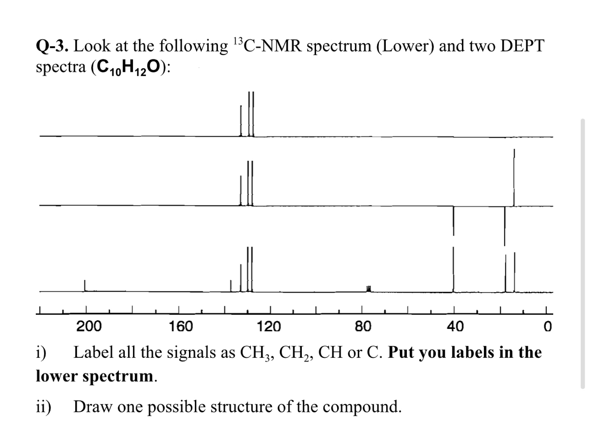
Answered: Q-3. Look at the following C-NMR… | bartleby
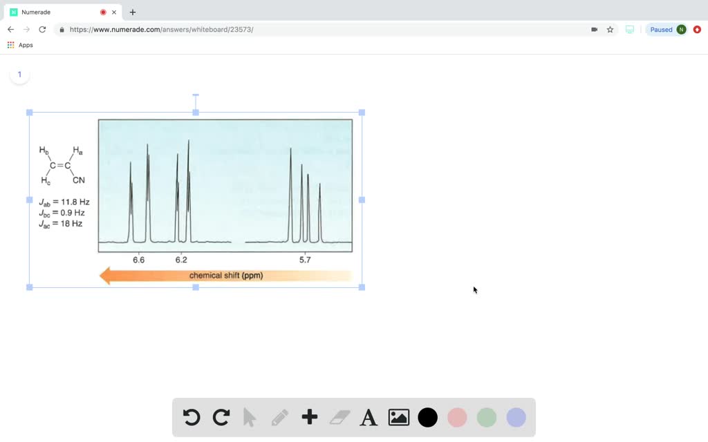
Label the signals due to Ha, Hb, and Hc in the ^1H NMR spectrum of acrylonitrile (CH2= CHCN). Draw a splitting diagram for the absorption due to the Ha proton.

Predict the H'NMR spectrum for Methylbenzene and label the ...
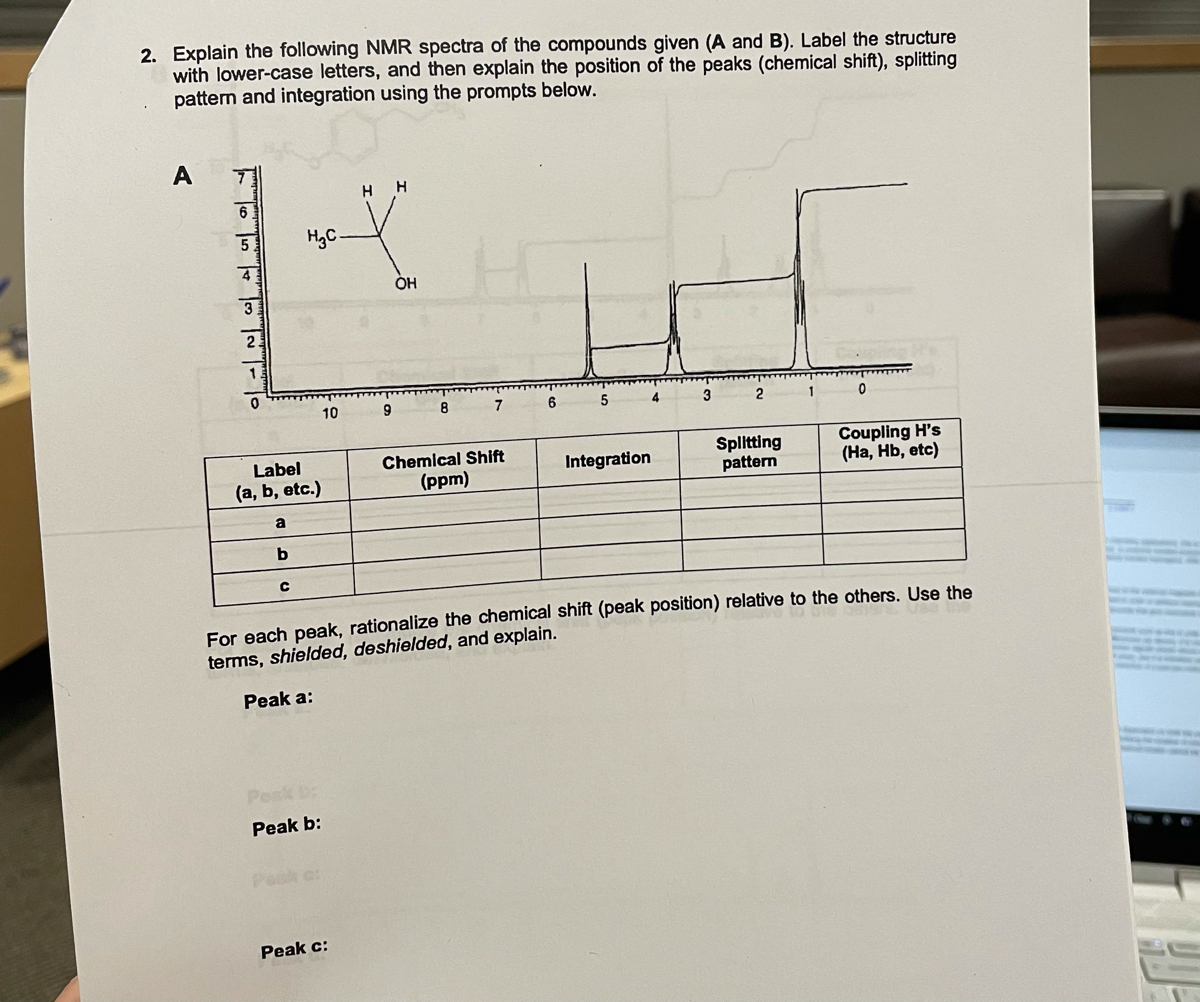
Answered: 2. Explain the following NMR spectra of… | bartleby
Proton NMR - How To Analyze The Peaks Of H-NMR Spectroscopy
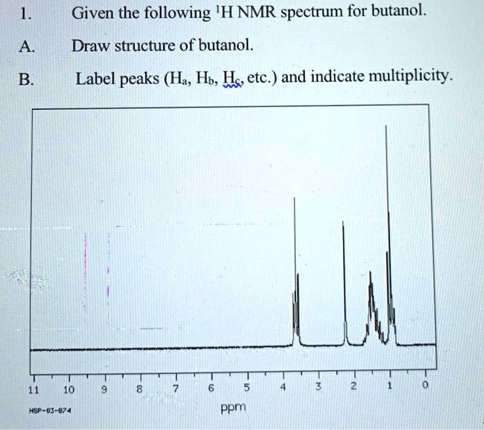
SOLVED: Given the following 'H NMR spectrum for butanol. Draw ...

Proton NMR practice 1
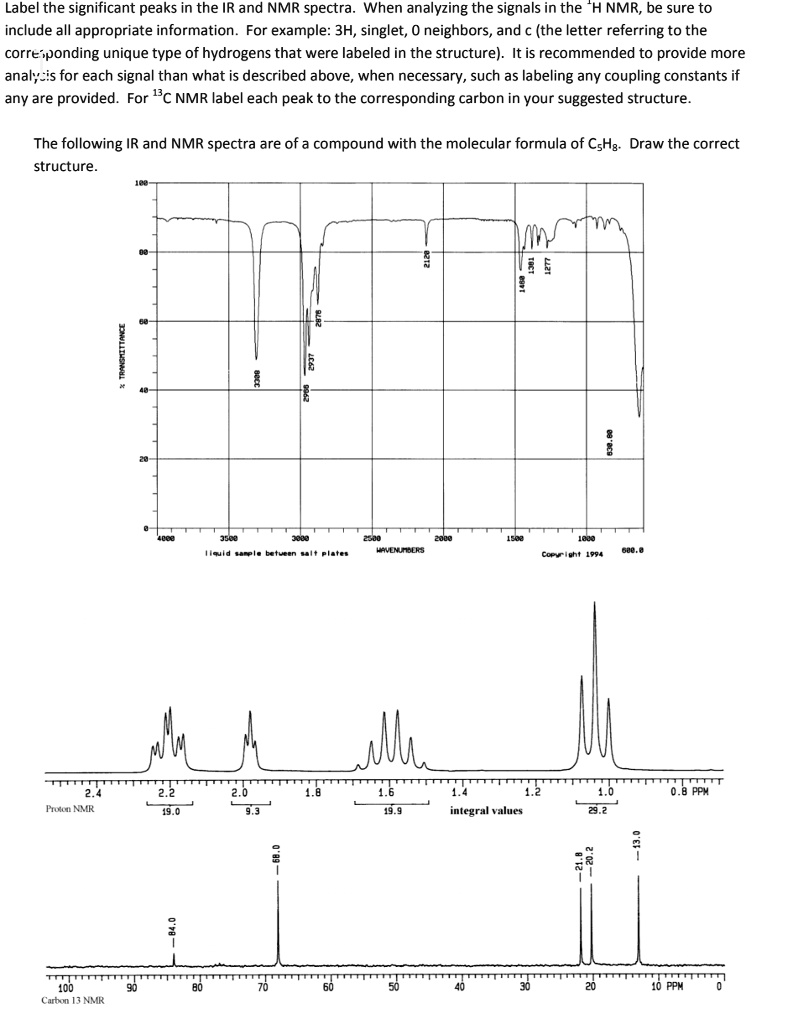
SOLVED: Label the significant peaks in the IR and NMR spectra ...

4.7: NMR Spectroscopy - Chemistry LibreTexts

NMR = Nuclear Magnetic Resonance - ppt video online download
Carbon-13 NMR Spectroscopy

Q.21558-14-45P Question: Label the signals due ... [FREE ...
6.6 ¹H NMR Spectra and Interpretation (Part I) – Organic ...
Post a Comment for "41 how to label nmr spectra"