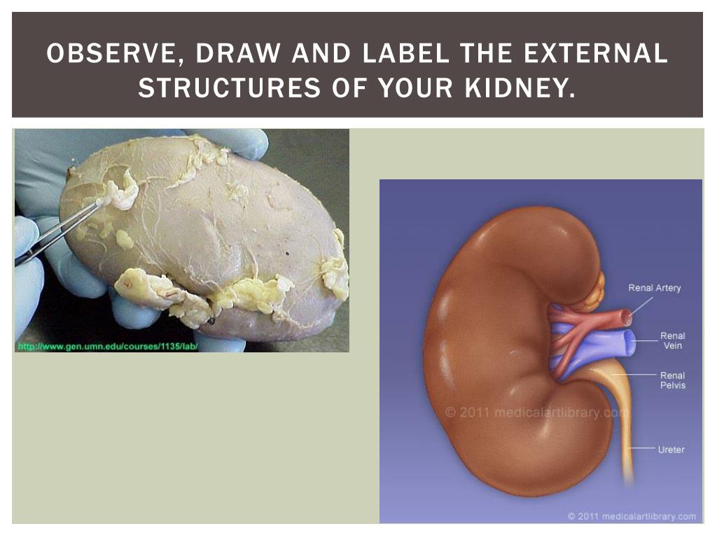
38 label the structures of the kidney
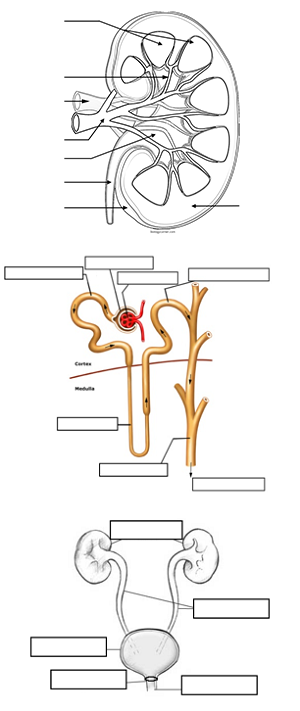
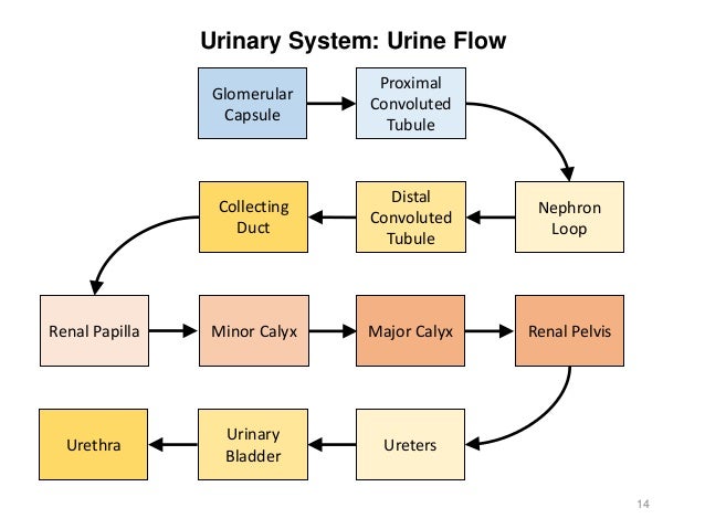
Urinary System Structures - Visible Body The kidneys, ureters, bladder, and urethra are the primary structures of the urinary system. They filter blood and remove waste from the body in the form of urine. The size and position of lower urinary structures vary with male and female anatomy. 1. Kidneys Filter Blood at the Top of the Urinary System (Get Answer) - Label the structures of the kidney. Label the structures ... Exercise 2: Internal Structures of the Kidney 1. Label the internal kidney structures in Figure 11.2 by filling in the blanks following the fig ure, 2. Label the path of urine drainage starting with the collecting ducts. Collecting dust papillary...
Urinary System - Label the Kidney and Nephron Students drag labels to the structures on the slide. Also, the diagram shows the relationship between the aorta, vena cava, and the renal vessels. While these aren't part of the urinary system, they are important in the physiology of the kidney. On the second slide, viewers see a close-up of a kidney that's been cut to show the internal structures.
Label the structures of the kidney
Kidney: Function and Anatomy, Diagram, Conditions, and Health Tips The kidneys are two bean-shaped organs in the renal system. They help the body pass waste as urine. They also help filter blood before sending it back to the heart. The kidneys perform many ... Lab 9: Exercise 40: Urinary System Flashcards - Quizlet Identify the structures of a kidney in the micrograph section of the renal cortex by clicking and dragging the labels to the correct location. Label the midsagittal male pelvis using the hints provided. ... Label the structures of the urinary system using the hints provided. A&P II (Ch 24 - 25) Flashcards | Quizlet nephron consists of renal corpuscle and renal tubule T/F: The juxtaglomerular apparatus is a structure of the nephron where the DCT contacts the afferent arteriole. true Label the structures of a nephron in the figure. Correctly label the following components of the urinary system.
Label the structures of the kidney. The Kidneys - Position - Structure - Vasculature - TeachMeAnatomy Internally, the kidneys have an intricate and unique structure. The renal parenchyma can be divided into two main areas - the outer cortex and inner medulla.The cortex extends into the medulla, dividing it into triangular shapes - these are known as renal pyramids.. The apex of a renal pyramid is called a renal papilla.Each renal papilla is associated with a structure known as the minor ... Kidney - Anatomy, Structure, Functions, Diseases and Treatments Anatomy of the Kidney The parenchyma is called the functional substance of the kidney. The parenchyma is divided into two major structures which are the outer renal cortex and the inner renal medulla. The renal cortex and the renal medulla take the shape of eight to eighteen cone-shaped renal lobes. automatic kidney segmentation, reconstruction, preoperative ... από Σ Ζάγκου · 2021 — Overall, it is obvious that 3D printed anatomical kidney models ... Table 3:Different label values for the different structures in KiTS19 labelmaps.121 σελίδες A&P 139 Urinary System Flashcards | Quizlet Label the structures of the male urinary tract. paired kidneys, paired ureters, a bladder and a urethra. The organs of the urinary system are outer fibrous coat, middle muscular coat, inner mucous coat. The layers of a ureter are Complete the sentences describing the functions of the kidneys.
προγραμμα αναπτυξης κοιτασματος δυτικου κατακολου (α ... organ(s) based on animal data: - Liver - Kidney - Lung ... Cool surrounding equipment, fire-exposed containers and structures with water.328 σελίδες Label the diagram (kidney internal structure): | Study.com The kidneys are the bean-shaped structures that are present between the last thoracic and third lumbar vertebra. They are situated dorsally to the inner wall of the abdominal cavity. A pair of... Kidney-Structure, Anatomy and Function - Online Biology Notes Kidney-Structure, Anatomy and Function Gross Structure Kidneys are bean-shaped organs, about 11 cm long, 6 cm wide, 3 cm thick and weigh 150 g. They are embedded in, and held in position by, a mass of adipose tissue. Each kidney is enclosed by a thin tough fibrous connective tissue called renal capsule that protects it from infections and injuries. Structure of the Kidney (With Diagram) | Organs | Human Physiology Nephron is the basic unit of kidney. The minute structure of the kidney is composed of a number of nephrons. Each human kidney possesses about 1 -2 millions of nephrons. Each nephron is made up of two main parts: (1) Malpighian Body, (2) Renal tubule. (C) Blood Vessels: The two important blood vessels of the kidney are: (1) Renal Artery (2) Renal Vein.
A&P 139 Urinary System Flashcards | Quizlet Label the structures of the male urinary tract. paired kidneys, paired ureters, a bladder and a urethra. The organs of the urinary system are. outer fibrous coat, middle muscular coat, inner mucous coat. The layers of a ureter are. Complete the sentences describing the functions of the kidneys. Kidney Anatomy, Parts & Function, Renal Cortex, Capsule, Nephron, Calyx ... The kidneys are highly vascular (contain a lot of blood vessels) and are divided into three main regions: the renal cortex (outer region which contains about 1.25 million renal tubules), renal medulla (middle region which acts as a collecting chamber), and renal pelvis (inner region which receives urine through the major calyces). Kidney - Structure, Different Parts and Functions - VEDANTU To summarize the structure of kidney diagram, the renal cortex comprises the outer part of this organ where the Malpighian corpuscles and the convoluted tubules of the nephrons exist. It is surrounded by fatty tissue at the outer portion for shock absorption and protection. The renal medulla is the part where the Henle's loop of all the nephrons lies. It also contains the renal pyramids of the kidneys. 25.1 Internal and External Anatomy of the Kidney As noted previously, the structure of the kidney is divided into two principle regions—the peripheral rim of cortex and the central medulla. The two kidneys receive about 25 percent of cardiac output. They are protected in the retroperitoneal space by the renal fat pad and overlying ribs and muscle.
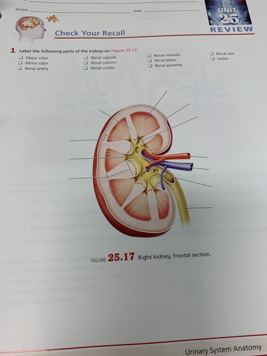
Solved: Section 25 UNIT 25 REVIEW Check Your Recall 1 Labe... | Chegg.com
Labeled Diagram of the Human Kidney - Bodytomy The renal medulla comprises a set of 8-18 conical structures called renal pyramids that are surrounded by the cortex. Portions of the cortex between two adjacent pyramids are termed as renal columns. Spread in these pyramids and the cortex, are the functional units callednephrons. The actual filtration of blood occurs in the nephrons.
Urinary System Review Guide
Μπούνιας-Διπλωματική-GR-EN.pdf - Artemis 13 Οκτ 2020 — structures: significantly improved efficiency and reliability,” ... “The KiTS19 Challenge Data: 300 Kidney Tumor Cases with Clinical.128 σελίδες
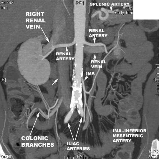
PA view Reconstructed image Origin and course of renal Arteries
The Structure and Function of the Kidneys - Verywell Health A pair of bean-shaped organs, the kidneys sit in the flanks, closer to the spine than to your belly. They are located just underneath your diaphragm and rib cage. They normally range in size from 8 to 14 centimeters (or 3 to 5.5 inches). Each kidney weighs between 120 grams (about a quarter-pound) to 170 grams (0.4 lbs).
Color and Label the Nephron
Solved Label the internal structures of the kidney in the - Chegg Label the internal structures of the kidney in the figure below using the terms: renal artery, renal vein, renal cortex, renal capsule, collecting duct, renal corpuscle, renal medulla, medullary pyramid, minor calyx, major calyx, renal pelvis, renal column, ureter. Show transcribed image text Expert Answer 100% (1 rating)
PPT - Kidney Dissection PowerPoint Presentation - ID:2118793
Solved Label the structures of the kidney. Renal Pelvis | Chegg.com The anatomy of kidney shows two regions. The outer region is called renal cortex and the inner dark r … View the full answer Transcribed image text: Label the structures of the kidney. Renal Pelvis Major Calyx Renal Cortex Minor Calyx Ureter Major Calyx Renal Cortex Renal Pelvis Minor Calyx Ureter Renal Pyramid Renal Medulla

Kidney Diagram In Human Body — UNTPIKAPPS
Kidneys: Anatomy, function and internal structure | Kenhub The kidneys are bilateral organs placed retroperitoneally in the upper left and right abdominal quadrants and are part of the urinary system. Their shape resembles a bean, where we can describe the superior and inferior poles, as well as the major convexity pointed laterally, and the minor concavity pointed medially.

Kidney Tubule Under Microscope - kidneyoi
Urinary system quizzes and labeled diagrams | Kenhub This gives you the opportunity to get a general feel of the appearance of each structure and their relations to the structures around them. Take a look at the urinary system diagram labeled below. You'll notice familiar structures like the bladder and ureters, as well as perhaps less familiar structures such as the renal artery and vein.
Activity 12-urinary-reproductive
The kidneys - The role of the kidneys in homeostasis - WJEC - GCSE ... The kidney. This diagram shows where the renal artery enters the kidney, and where the renal vein leaves. The kidney is packed with around a million structures called nephrons. These nephrons ...

Nephron Illustration Part Of The Kidney Stock Illustration - Download ...
Kidney structure (anatomy) - SlideShare Kidney structure (anatomy) Dec. 14, 2014. • 56 likes • 48,180 views. D.A.B.M. Download Now.
Post a Comment for "38 label the structures of the kidney"